Published on 24 Sep 2025 by rovertech
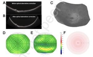
Methods: In this cross-sectional study of 147 adults (51 HM, 96 controls), ultra-widefield swept10 source optical coherence tomography (SS-OCT) with Gaussian curvature mapping quantified RC across six concentric zones (diameters 1-15 mm). Ocular parameters, including Corneal K, lens thickness (LT) and axial length (AL) were measured using Lenstar LS 900, and retinal thickness (RT), choroidal thickness (ChT), choroidal vascularity index (CVI), and RC were measured by ultra-wide field SS-OCT. Correlation and regression analyses identified factors influencing RC.
Results: HM participants had longer AL, thinner ChT, higher CVI, and flatter macular RC versus NHM 16 group (p<0.05). Multiple linear regression analysis demonstrated that peripheral retinal regions beyond 6 mm exhibited significant negative correlations with AL (r = -0.296 to -0.478), while central regions within 9 mm showed positive associations with CVI (r = 0.180-0.214). RT was negatively correlated with RC in perimacular regions (r = -0.19), but no significant association was observed between equivalent spherical refractive power (SER) and RC in any measurement region.
Conclusions: Retinal curvature alterations in high myopia demonstrate spatial heterogeneity, featuring peripheral biomechanical adaptation to ocular elongation and central structural changes modulated by choroidal vascular integrity. Integrating RC into clinical assessments could enhance monitoring of biomechanical changes in HM. Longitudinal studies are needed to validate its predictive role in myopia progression.