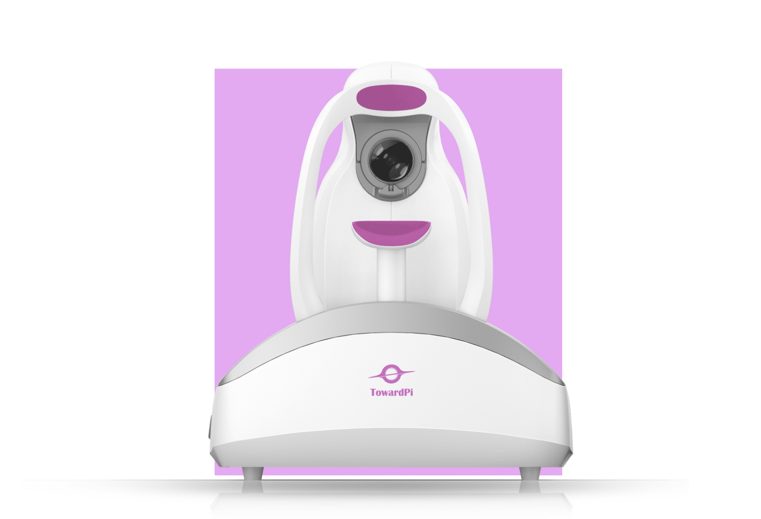
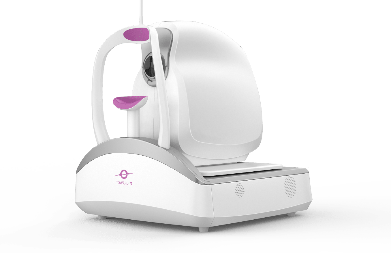
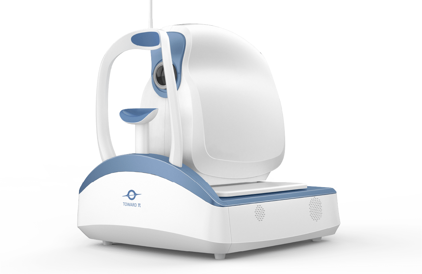
TOWARD PI®BMizar 400KHz Full-Range SS-OCT (BM-400Max)

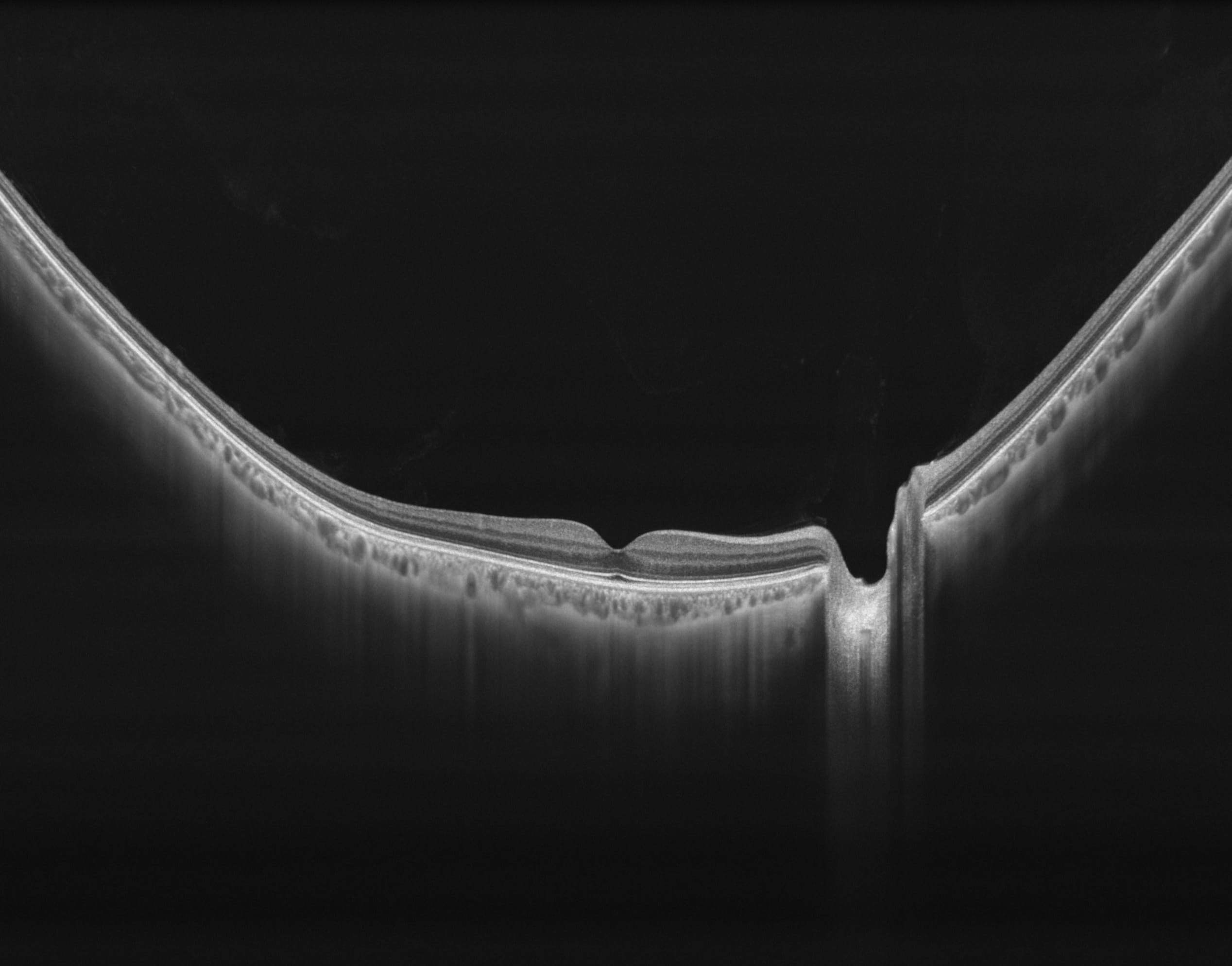
400kHz speed full-range swept-source OCT for anterior and posterior

  • 400,000 scans per second tdsafsadfdfo power your practice
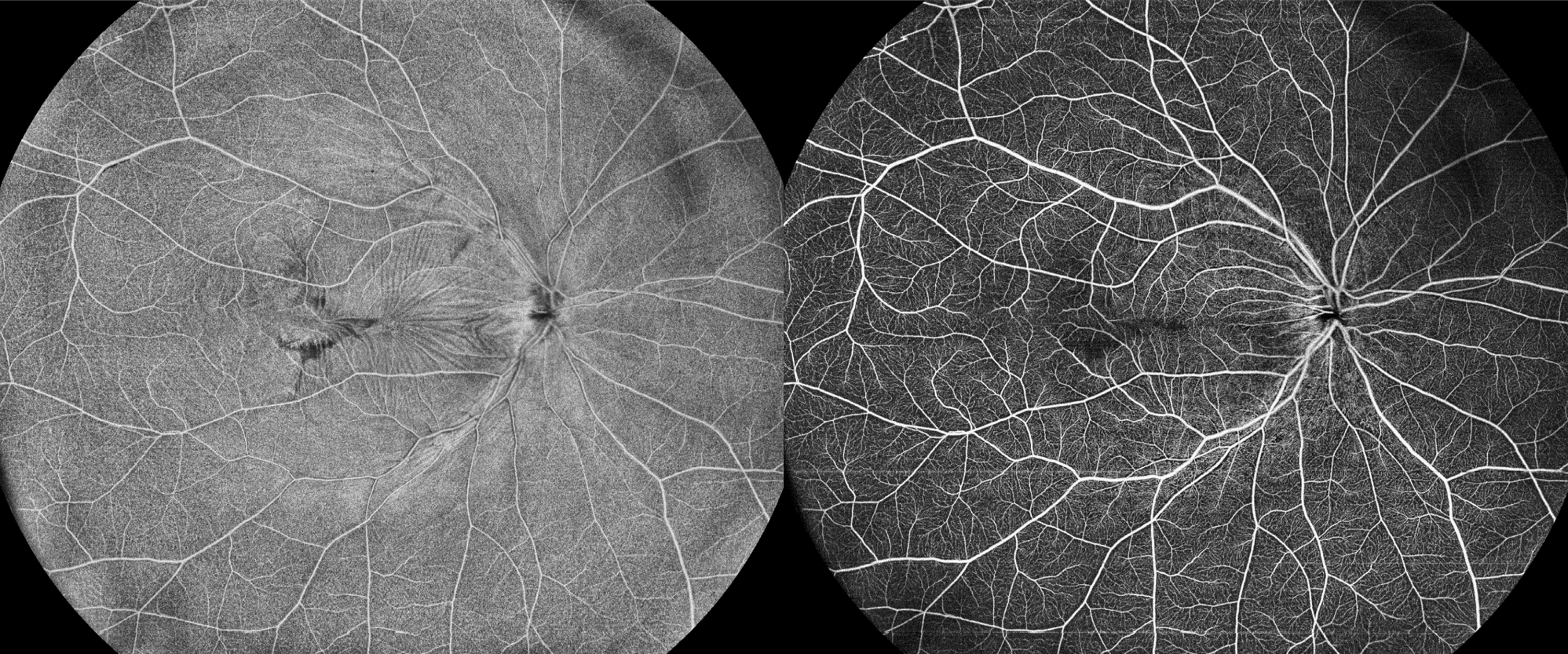
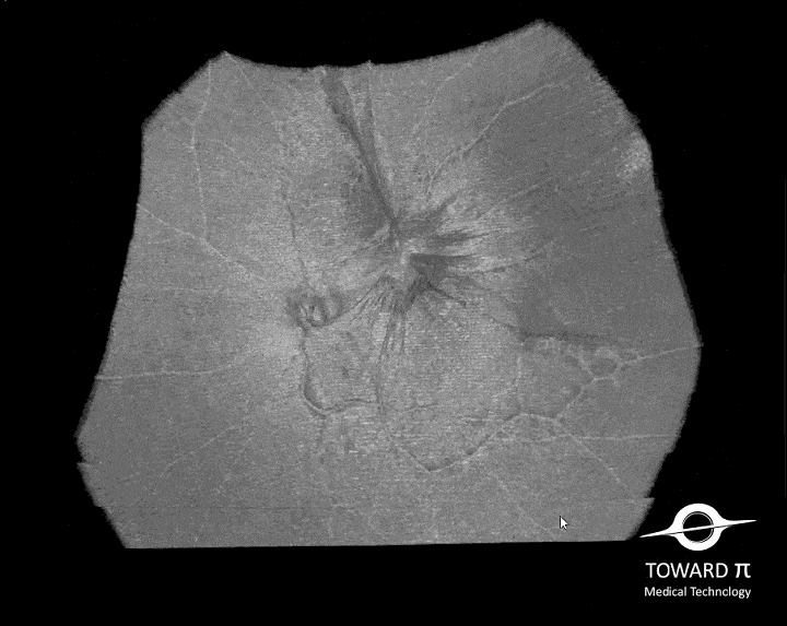
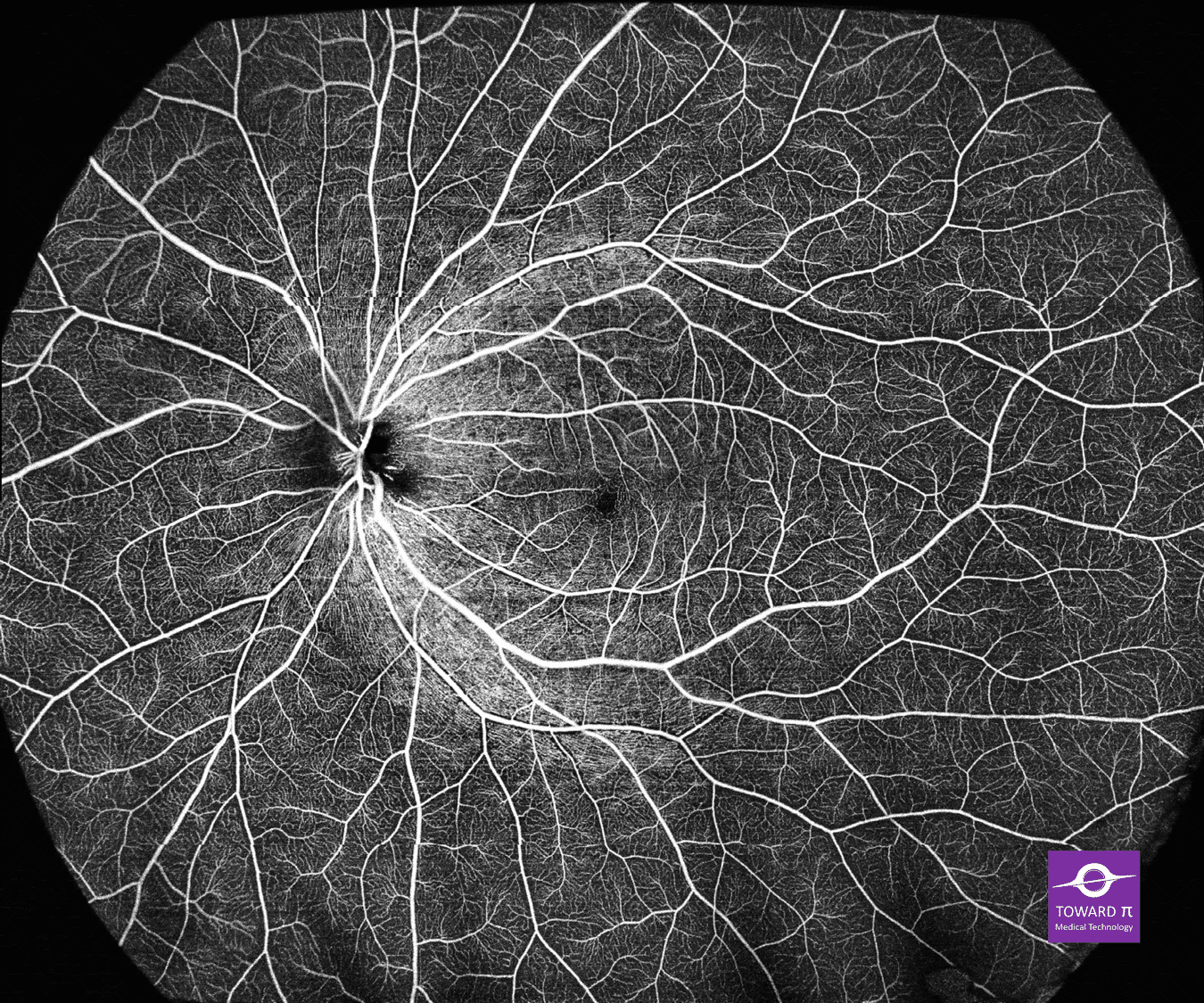
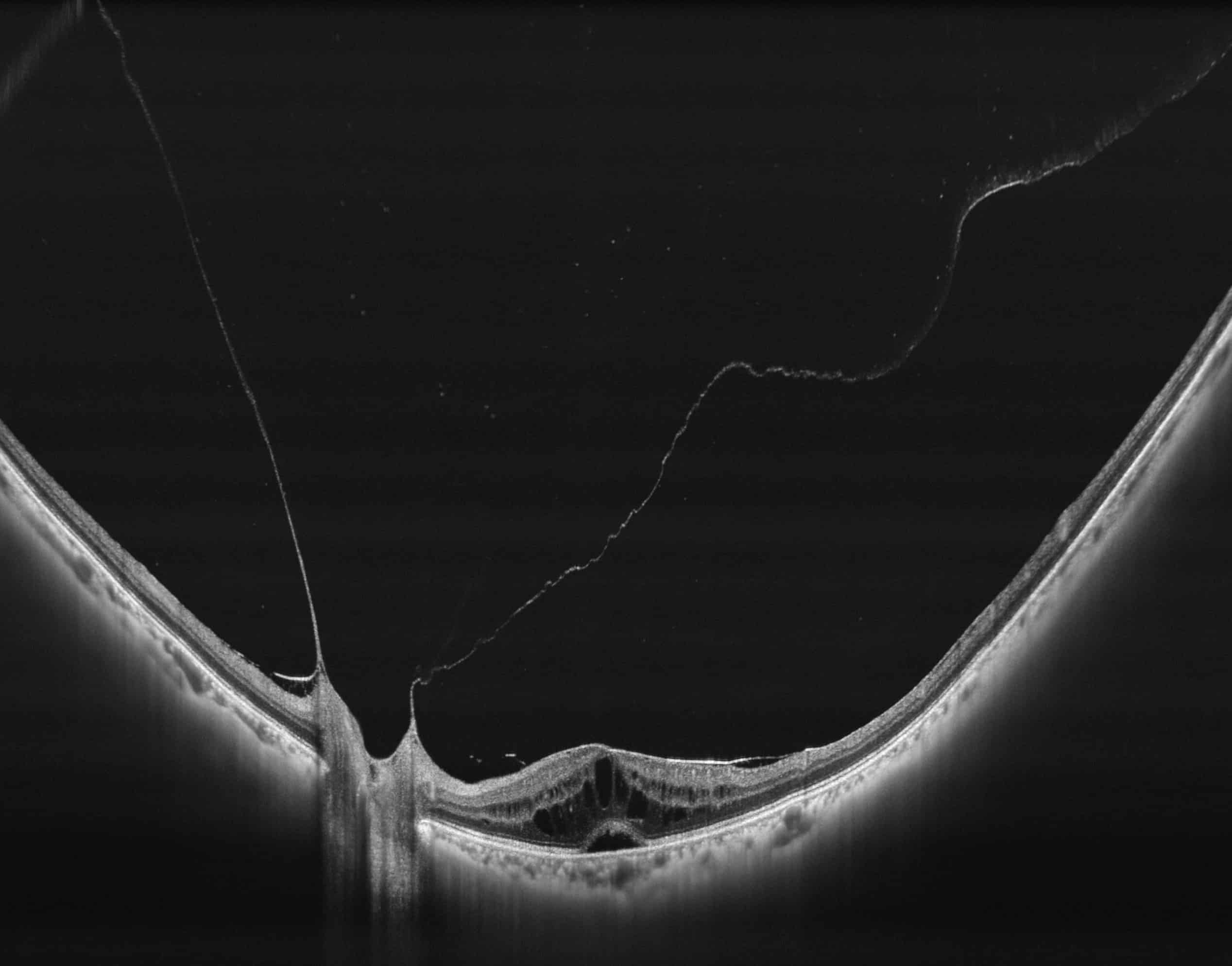
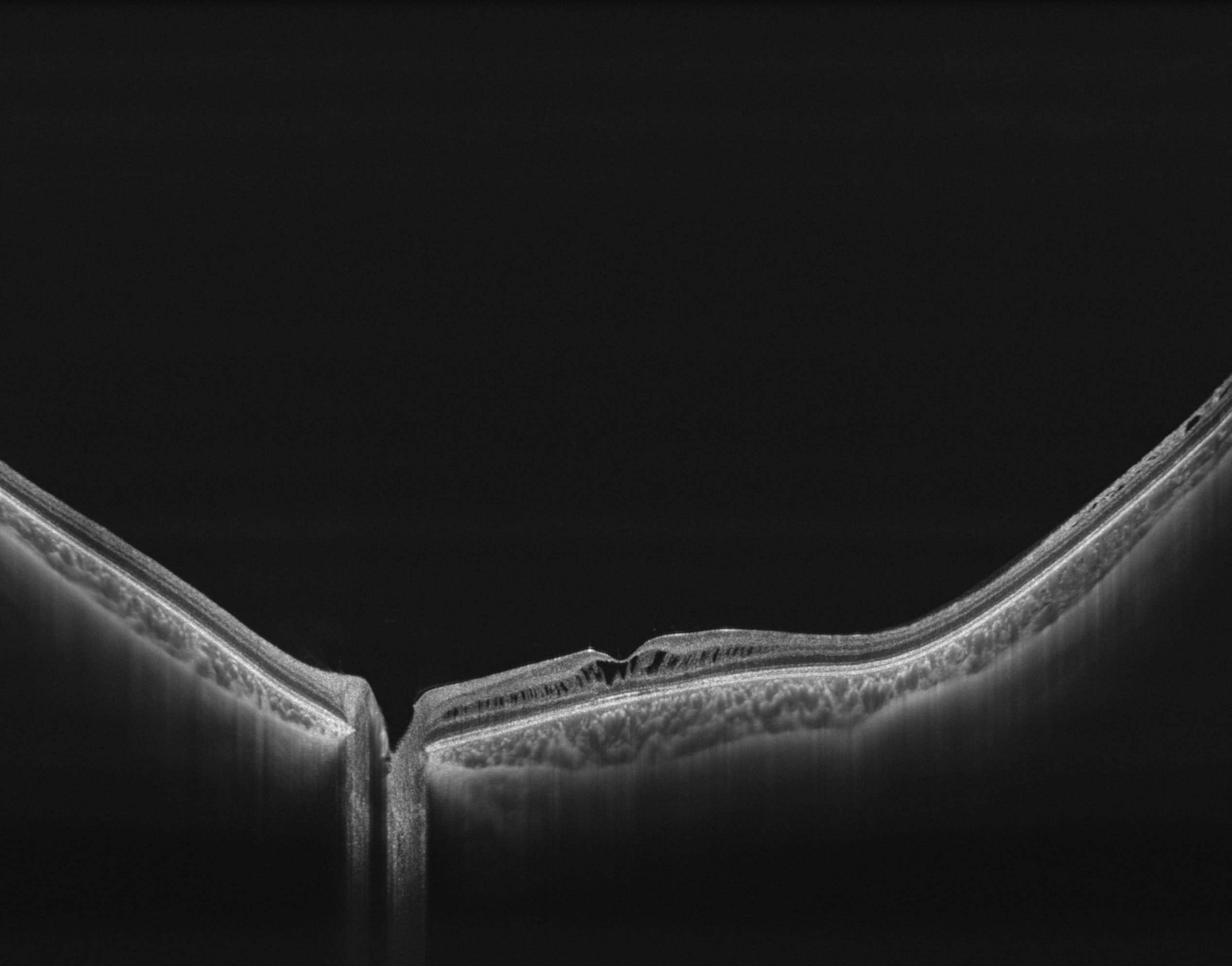
  • Full-Range wide-field Swept Source OCT
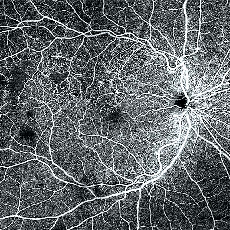
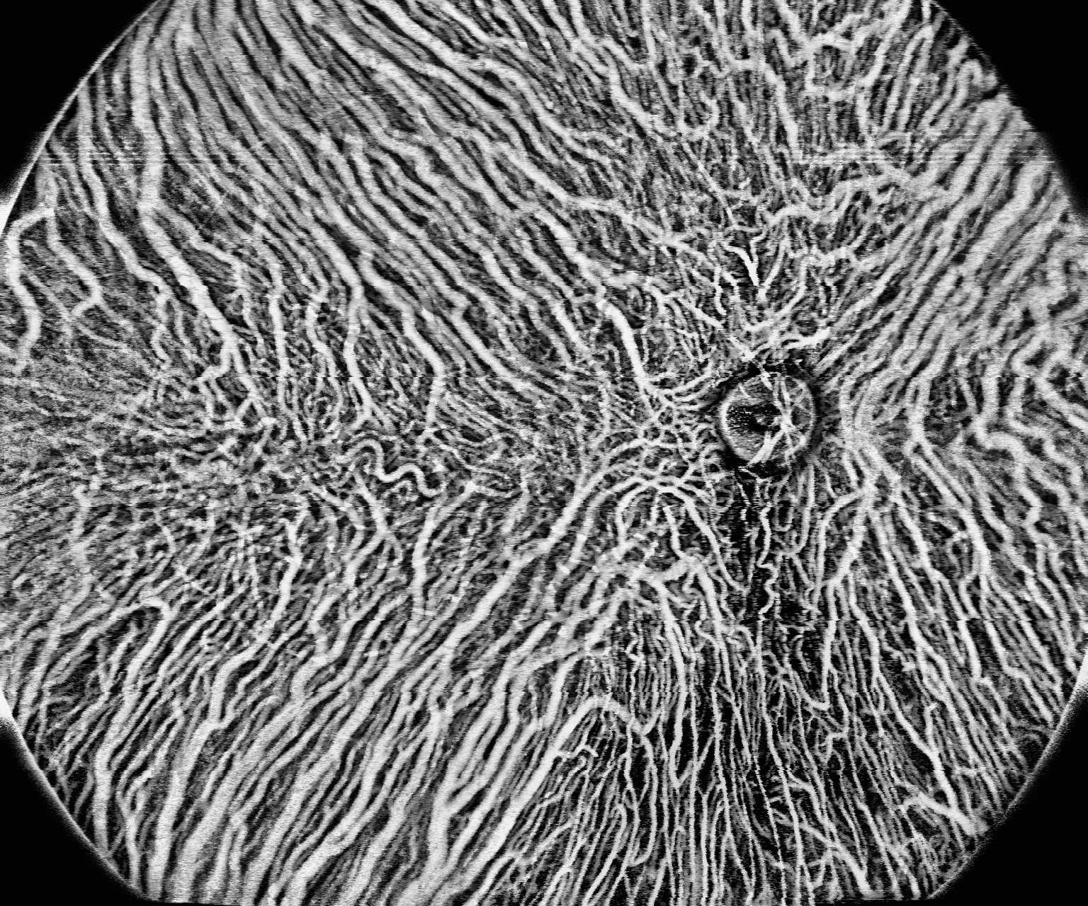
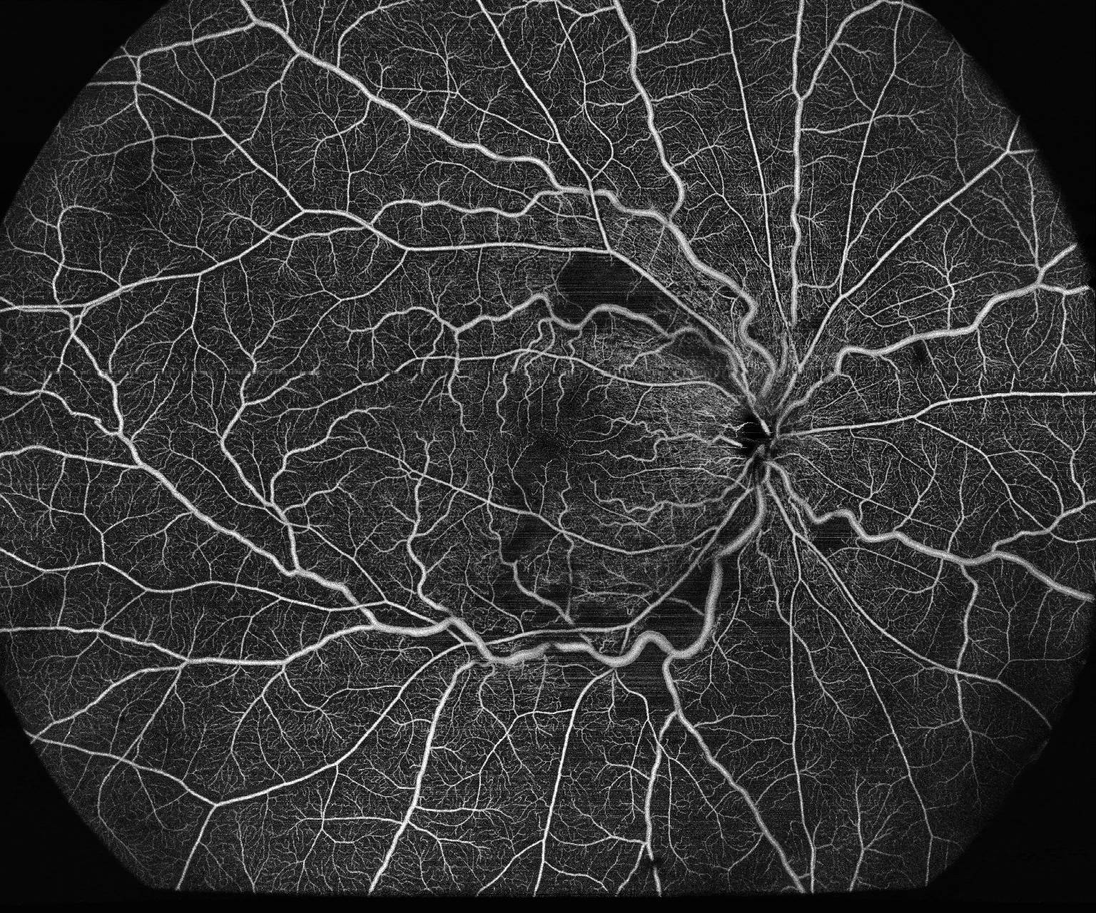
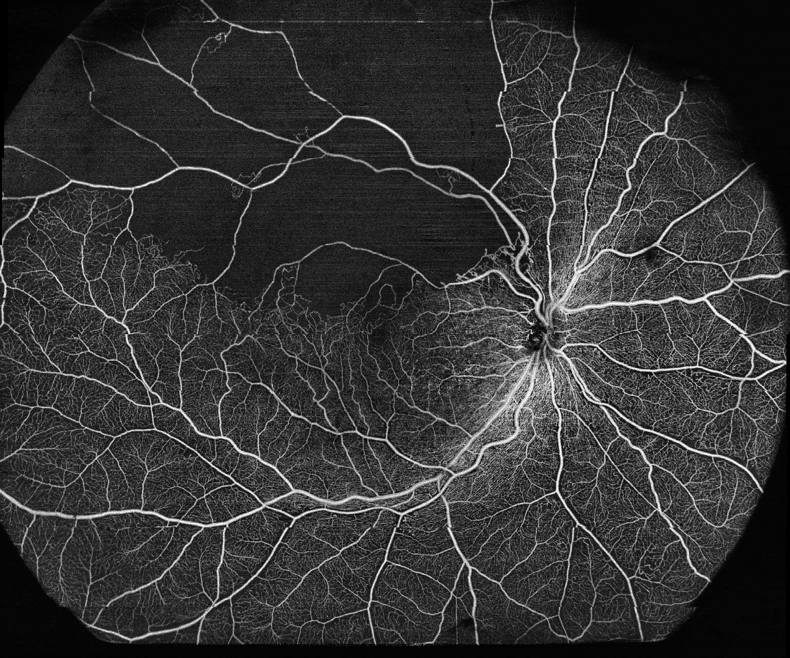
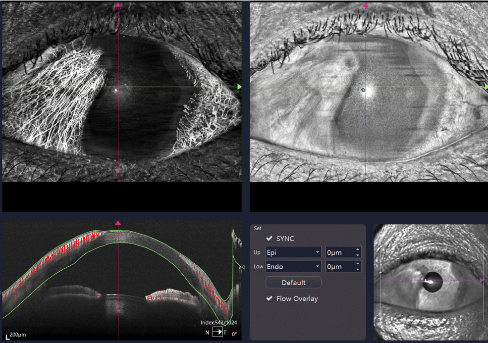
  • Wide-field OCTA
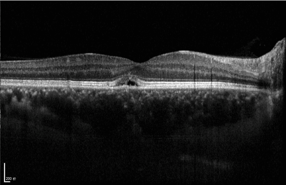
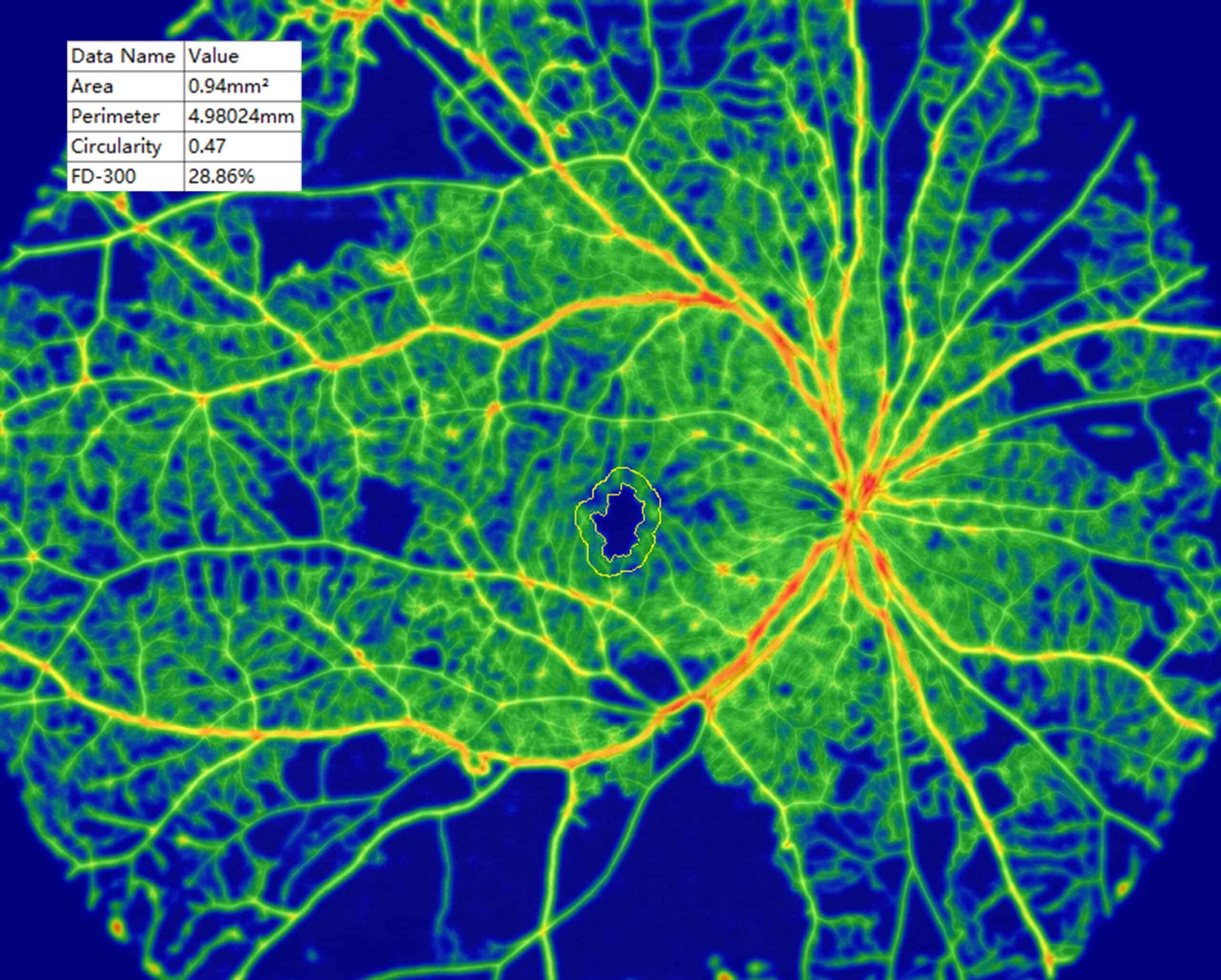
  • Choroid OCTA with Quantification Parameters
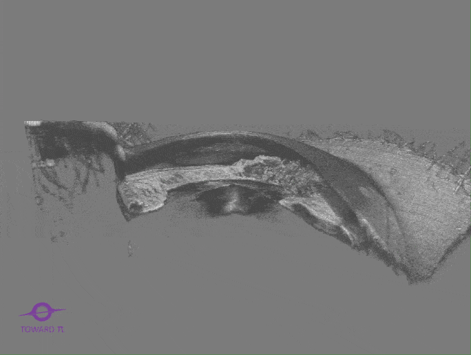
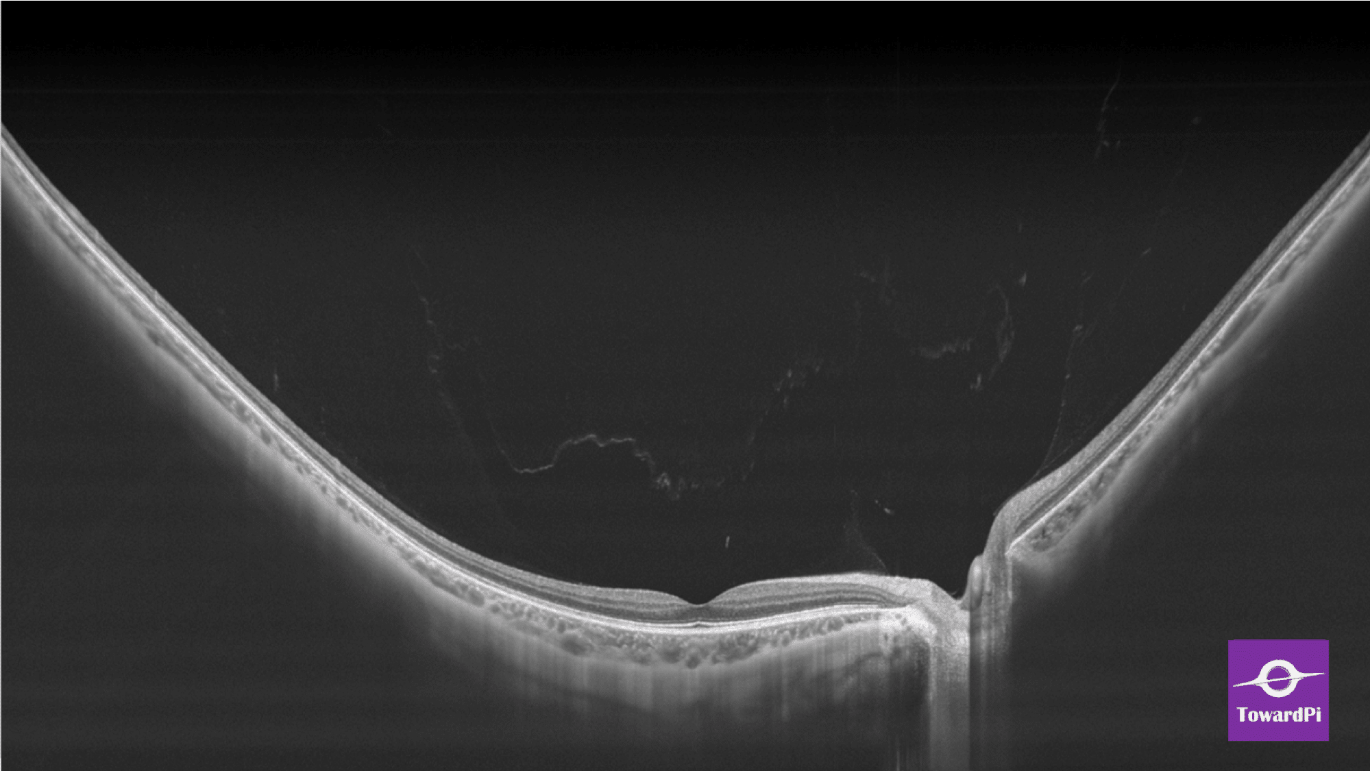
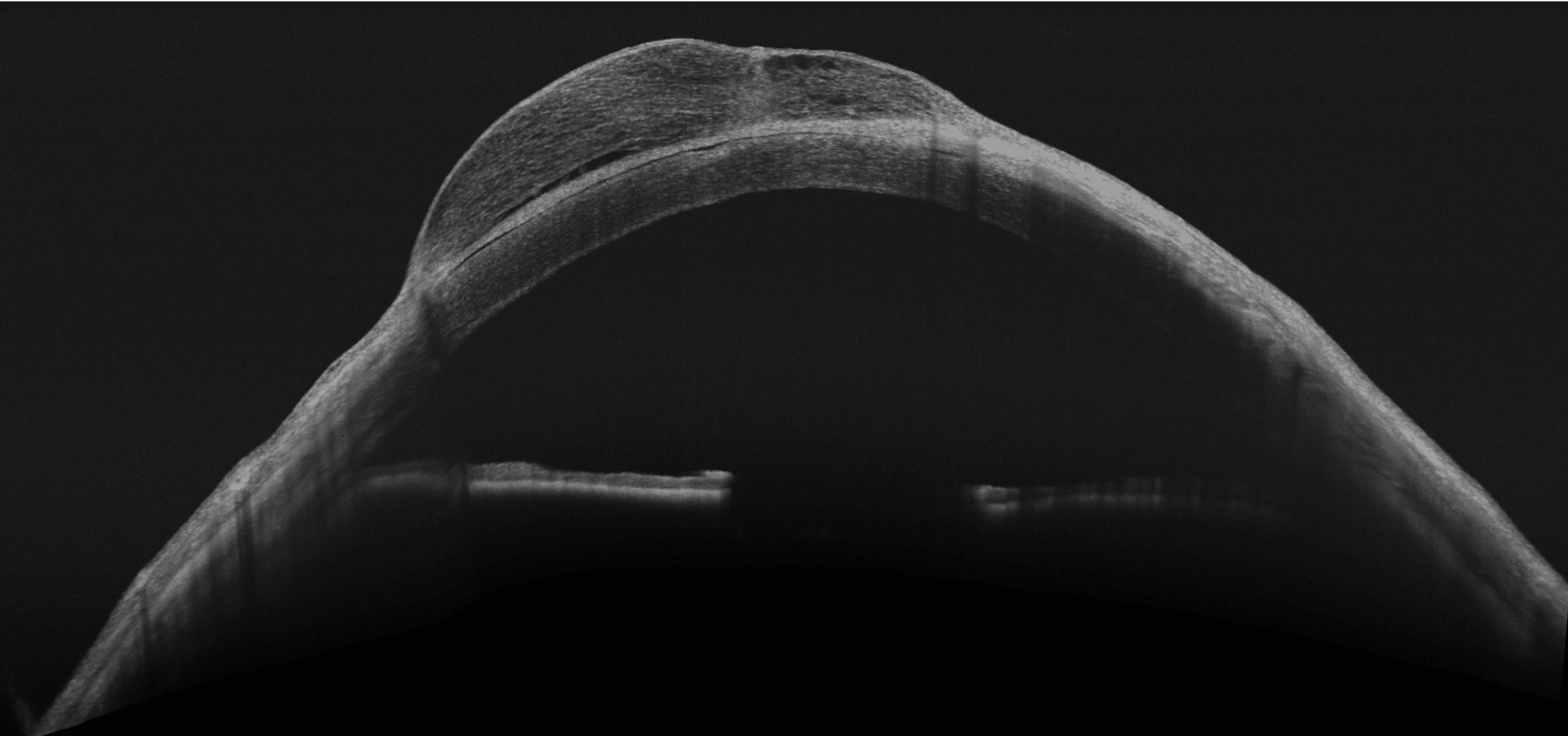
  • HD Anterior Scan with Anterior OCTA
Get a Quote
Product Details Image

The best of both worlds
The TowardPI® Swept-Source OCT

BMizar (BM-400K) showcases the industry’s most powerful OCT “engine” with a 400kHz swept-source. Its exceptional optic design elevates the experience of OCT Angiography to an unprecedented level. With high-definition imaging of the retina, choroid, and anterior, it effortlessly covers an incredibly large area in a single scan (achieving a 24 mm width OCTA in just 7-15 seconds).

This revolutionary technology is a game changer for clinical practices, particularly in the diagnosis and treatment of diseases such as DR, RVO, RAO, and more. Furthermore, the BMizar offers a vast dioptric adaptive range, making it feasible to perform all types of OCT/OCTA scans on small animals like rabbits, cavies, and even mice without the need for additional lenses.

It is truly a dream machine for any OCT user.

When it comes to precise diagnostics and successful treatment of the anterior segment of the eye, it provides the basis for a comprehensive and reliable diagnosis

  • 24 x 20 mm OCTA Wide-Field OCTA
  • In seconds: Retina OCT+OCTA in just one single measurement
  • High optical resolution of 3.8 µm of the retina
  • 10 Billion maximal voxels
  • In-built AI Segmentation
Download Brochure

Normal OCT just show 12x12mm Bmizar OCT show 24x20mm OCTA

Normal SD OCT systems typically offer a maximum scan range of 6mm to 12mm for posterior segment imaging and up to 15mm axial depth in advanced swept-source variants, constrained by speed and resolution limits.
In contrast, the BMizar OCT, a high-speed 400kHz full-range swept-source system, achieves an expansive maximum scan range of 24mm x 20mm for ultra - widefield posterior OCTA and 24mm length for anterior segment scan, enabling comprehensive coverage of the retina and choroid in a single acquisition. this advancement significantly enhance diagnostic efficiency for conditions like diabetic retinopathy.

Product Details Image
Top: Normal OCTA 12mmx12mm
Left: Bmizar OCTA 24mmx20mm

Revolutionizing Retina and Anterior Precision

Structural en face OCT and Anterior segment helps in evaluating the topographic location of the lesions.
Epiretinal membrane 3D / Iridoschisis 3D

Product Details Image

How deep and how wide we can go

Here’s CSR(Central serous chorioretinopathy) case. In SD-OCT, the outer limit of the choroid and the sclera cannot be reliably identified due to the scattering of light from RPE. SS-OCT has enabled better visualization of choroidal structures and choroidal-scleral interface, which is useful for the diseases of pachychoroid. The TowardPI ® BMizar uses Swept-Source OCT with Full-range scanning technology to provide 3.8 μmresolution and 1000-1300 Wavelength of retina structures that conventional systems cannot achieve. While SD OCT + EDI systems offer high scan depth, they are limited in resolution to approximately 5-8 μm and 850±20nm wavelength , insufficient for revealing fine details. We offer

TowardPI® BMizar
Resolution Box Image Equals to 120 degrees fundus,24mm HD line B-scan shows that the details of posterior segment structure imaging, including retina and choroid, PPVP (Posterior precortical vitreous pocket) above macula and Cloquet’s canal above optic disc were clearly visualized. and the most important is the image signal strengths are identical.
Normal SD-OCT
Resolution Box Image Normal SD-OCT

Images you must have seen

Full Range SS-OCT Images – impressive, convincing, simply sharp

Crystal Clear Image

Full Range retina vessels

Crystal Clear Image

Full Range Choroid vessels

Crystal Clear Image

Retinoschisis

Crystal Clear Image

Cystoid macular edema

Crystal Clear Image

Normal Eye (vitreous, retina and choroid)

Crystal Clear Image

Retinal Vasculitis

Crystal Clear Image

Branch Retinal Vein Occlusion

Crystal Clear Image

Flow density & FAZ indexes, Diabetic Retinopath

Crystal Clear Image

Hereditary Deep Dystrophy Of Cornea

Crystal Clear Image

Pterygium

How the TowardPi® Full-range SS-OCT works

Find out why the TowardPi® OCT technology produces significantly higher resolution images than SD-OCT.

Compare BMizar and Yalkaid

Best of both worlds
OCT and OCTA technology

  • A-scan Speed
  • Max. OCTA size
  • Max. Depth Anterior
  • Tracking speed
Product Details Image

BMizar (BM400K-MAX)

  • 400,000Hz
  • 24x20mm (HD)
  • 6mm
  • 128Hz
Product Details Image

Yalkaid (YG100K-MAX)

  • 100,000Hz
  • 15x15mm (HD)
  • 14mm
  • 100Hz

Technical Data

OCT Model:BM-400K

OCT optical source
Swept Source
Center wavelength
1060nm
Scan speed
400,000 A-scans/sec
Max. Length (posterior)
24mm
Scan depth (anterior)
6mm
Axial best digital resolution
1.9μm
Fundus Imaging Methodology
Scanning Laser Ophthalmoscopy (SLO)
Eye tracking speed
128Hz
SLO FOV
60° ×60°
Minimum pupil diameter
2.0mm
OCT Angiography Max. scan size (montage)
42mm×40mm
OCT Angiography Max. Single scan size (posterior)
24mm×20mm
Maximum resolution (single scan)
1536×1280
Software Functions

Look for reliable eye diagnostic instruments? Our team is here to help.

Get expert guidance and support to choose the right instruments for your practice.

Contact Us
This site is registered on wpml.org as a development site. Switch to a production site key to remove this banner.