Published on 28 Jun 2026 by admin website
Retinal curvature remodeling following PSR in high myopia quantified by UWF full range SS-OCTA!
Research recently published in <Photodiagnosis and Photodynamic Therapy> quantitatively assessed retinal curvature (RC) and microvascular changes following posterior scleral reinforcement (PSR) by using UWF full range SS-OCTA!
After PSR surgery, all 21 enrolled high-myopic eyes exhibited significant axial shortening and progressive retinal flattening with a consistent decline in mean RC. Notably, a significant correlation between changes in RC and axial length (AL) emerged as early as 1 month postoperatively, suggesting that RC may serve as a sensitive early biomarker for treatment efficacy. Regional alterations in RC demonstrated significant associations with retinal and choroidal flow changes, indicating that mid-peripheral flattening was associated with higher choroidal blood flow, providing evidence for the structural and hemodynamic remodeling following PSR.
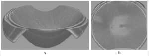
A 400kHz speed swept source OCTA BMizar, TowardPi Medical was used in this study to acquire 24mmx20mm UWF OCTA images for retinal curvature according to AL with AI-based algorithm. A curvature map detailing 3-dimension Gaussian curvature of each point is generated, providing a quantitative indicator of eyeball morphology for long-term clinical monitoring and quantitative evaluation of surgical outcome.
This research is authored by Dr. Xiaoxiao Wu, Dr. Xiyue Tan, etc. team of Prof. Junguo Duan from Chengdu University of Traditional Chinese Medicine.
Link to original text:
https://lnkd.in/d-jW7HrT
One of 11 publications on Retinal Curvature using TowardPi — Academically repeatedly verified OCT quantification!
#TowardPi HongKong