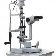
The OCT for general screening
The Mocean 3000 Plus achieves the optimum balance between cost and performance with its fundus surface imaging system. It has been developed for screening in general eye clinics.
OCT with Real- time widefield LSO image
The Mocean 3000 Plus equipped with LSO ( Line Scanning Ophthalmoscopy), and provides simulataneously high quality fundus image, which is easy for physicians to localize to lesion.


Wide area scan (6 x 6 mm / 12 x 12 mm)
The 6 x 6 mm/ 12 x 12 mm wide area map enables analysis of [NFL+GCL+IPL] status in and around macula, around optic disc, and even in the peripheral area.

*OCT Scan length can be switched between 6mm and 12mm
High-speed scan (Max. 36,000 A-scans / s)
The high-speed scan (Max.36,000 A-scans / s) and high-speed averaging (Max. 50 images) achieve high-definition B-scan image.

Ease of Use for Capturing Small Pupils
OCT-LFV image, which is a live projection image with reflection at retina, will show the live Fundus image clearly even in cases of smal pupilis. Disc, retinal vesseles and scanning position is very easy to see.
Complete OCT Functionalities
- For Macula
– Line Scan
– 3D Macula Cube Analysis
– Six LineRadial Scan
- For Glaucoma
– Wide Scan (12 mm × 12 mm)
– Macula(V) Glaucoma Analysis
– 3D Disc Analysis
- For Anterior Segment– Anterior HD Line– Anterior Six-Line Radial

Multifunctional follow-up
The multifunctional follow-up allows analysis of all the data obtained with the OCT and detailed observation of choronological change in retinal thickness and status. This function displays progression of pathology over time. The comparison mode displays two images selected by the user.

Progression display
Customized report
The layout of the reports can be customized and the data from separate reports of each scan pattern can be summarized in a single report to avoid printing multiple pages.

Video


