Published on 10 Jul 2026 by admin website
is a common eye condition affecting the macula — the central part of the retina responsible for sharp, detailed central vision. It is a leading cause of central vision loss in people over 50, particularly in aging populations like Hong Kong.
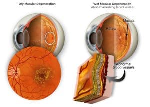
Dry (Non-exudative / Atrophic) AMD: Accounts for 85-90% of cases. Yellow deposits called drusen accumulate under the macula, leading to gradual thinning and atrophy of the retinal pigment epithelium (RPE) and photoreceptors. It progresses slowly but can lead to geographic atrophy (GA).
Wet (Exudative / Neovascular) AMD: Accounts for 10-15% but causes 80-90% of severe vision loss. Abnormal new blood vessels (choroidal neovascularization, CNV) grow from the choroid, leaking fluid or blood and causing rapid damage and scarring. Dry AMD can convert to wet.
Other forms of maculopathy exist (e.g., central serous chorioretinopathy, myopic maculopathy), but AMD is the most common age-related type.

Early stages may be asymptomatic — regular screening is essential. Common signs include:
Blurred or reduced central vision (difficulty reading or recognizing faces).

Metamorphopsia (visual distortion): Straight lines (door frames, grids) appear wavy, bent, or broken — a hallmark symptom.
Central scotoma (dark or blank spots in central vision).
Faded color perception or need for brighter light when reading.
Often affects one or both eyes; wet AMD can worsen suddenly.
Self-monitoring: Use an Amsler Grid — cover one eye, focus on the center dot, and check for distortions.
Primary: Aging (risk rises sharply after 50), genetics (family history, complement factor H variants).
Modifiable: Smoking (2-4x higher risk), hypertension, high cholesterol, cardiovascular disease, obesity, prolonged UV exposure, high-fat diet.
More common in Caucasians for early forms, but affects all ethnicities.
Visual acuity test + Amsler Grid.
Dilated fundus examination (observe drusen, hemorrhage).
Detects drusen, RPE changes, fluid, CNV, and atrophy. Excellent for diagnosis, monitoring treatment response, and follow-up (devices like TowardPi, Optovue, Zeiss, Nidek are widely used).
OCTA (OCT Angiography): Visualizes blood flow without dye.
Fundus Fluorescein Angiography (FFA) / Indocyanine Green Angiography (ICGA): Confirms neovascularization.
Color fundus photography for documentation.
OCT is indispensable in modern AMD management for early detection and personalized care.
Dry AMD: No cure. AREDS2 nutritional supplements (lutein, zeaxanthin, vitamins C/E, zinc) can slow progression in intermediate/late stages. Lifestyle: Quit smoking, eat leafy greens/fish, UV protection, regular eye exams.
Wet AMD: Intravitreal anti-VEGF injections (e.g., Ranibizumab/Lucentis, Aflibercept/Eylea, Faricimab/Vabysmo, Conbercept) are first-line. They inhibit abnormal vessel growth and leakage, preserving or improving vision. Typical regimen: monthly loading doses, then treat-and-extend.
Others: Photodynamic therapy (PDT), laser (less common), emerging therapies (gene therapy, new agents).
Advanced cases: Low-vision aids (magnifiers, adaptive devices).
Prognosis: Early intervention is key. Timely treatment for wet AMD controls progression effectively; geographic atrophy in dry AMD remains challenging.
黃斑病變(Macular Degeneration,主要指年齡相關性黃斑變性,Age-related Macular Degeneration, AMD) 是影響黃斑區(視網膜中央負責精細視力的部分)的常見眼疾,是50歲以上人士中心視力喪失的主要原因之一,尤其在香港及全球高齡人口中普遍。
1 OCT systems (e.g., TowardPi YAlkaid/BMizar/Zalioth series, Optovue, Nidek NFC700, Zeiss) are essential for AMD screening, AI-assisted analysis (macular thickness maps, RNFL), and patient data management in hospitals like Hong Kong Eye Hospital .
2 Highlight in proposals: Non-invasive, fast scanning, AI upgrades, data privacy (HK compliance), compatibility with existing systems.
3 Useful for clinician feedback collection, lease agreements (deposit, maintenance, ownership transfer), and marketing content emphasizing early AMD detection via OCT.