Published on 30 Jun 2026 by admin website
Your eye doctor will ask questions about your health and medical history and do a thorough examination of your eyes. These tests can show whether you have vision loss and what may be causing it. Tests may include:
1.Vision test. A vision test, also called a visual acuity test, uses eye charts to measure how well you can read a series of letters. One eye is tested at a time, while the other eye is covered. Your eye doctor determines how well you can see and what kind of corrective lenses you may need.
2.Microscopic exam. Your eye doctor may use a specialized microscope to look at the outer eye and structures inside the eye. This exam is often done with a device called a slit lamp. This uses bright light and a microscope to see precise details of structures or irregularities in your eye. Your eye doctor will look at the condition of the nerves at the back of the eye. This exam also can show if you have a cataract in a lens. With this exam, you may have eye drops to enlarge your pupils.
3. Intraocular pressure test. This measures the pressure inside your eye. There are multiple devices available to do this.
4.Questionnaire. You may fill out a form that asks you questions about how your vision affects your ability to do daily activities. If you have a cataract, this information helps in developing a treatment plan.
When your prescription glasses or contacts can’t clear your vision, the only effective treatment for cataracts is surgery.
Talk with your eye doctor about whether surgery is right for you. Most eye doctors suggest considering cataract surgery when your cataracts begin to affect your quality of life. This may include your ability to perform daily activities, such as reading or driving at night.
For most people, there is no rush to remove cataracts because they usually don’t harm the eyes. But cataracts can worsen faster in people with certain conditions. These include diabetes, high blood pressure or obesity.
Waiting to do cataract surgery typically won’t affect how well your vision recovers. Take time to consider the benefits and risks of cataract surgery with your healthcare team.
If you choose not to have cataract surgery now, your eye doctor may recommend periodic follow-up exams to see if your cataracts are getting worse. How often you’ll see your eye doctor depends on your situation.
The most common type of cataract surgery is called phacoemulsification (fak-o-e-mul-sih-fih-KAY-shun). During this process, a probe is inserted into the eye and uses ultrasound energy to rapidly move the tip of the probe back and forth to break up the cataract. The doctor then suctions out the lens, as seen in the top image. The outer housing of the lens, called the lens capsule, is left in place. After removing the lens, the doctor places the artificial lens into the capsule where the natural lens used to be, as seen in the bottom image.

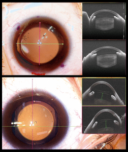
With the reliable real-time guidance of high-resolution SS-intraoperative OCT, the challenging pediatric cataract surgeries were performed with high-efficiency and high-precision by using digital navigation surgical microscope ( Towardpi Focean)
得益於Toward PI SS OCT的1060nm Wavelengh 可以穿透因白內障被乳化的晶體直達視網膜,因此醫生在評估白內障階段已經可以得知手術後預先得知是否會因為視網膜問題而導致視力下降,而非白內障本身。反之亦然,然而在白內障手術過程中醫生亦可以通過TowardPi Focean 手術顯微鏡動態地觀察手術中的每一個步驟,這樣對優化手術以及未來對眼科醫生的培訓帶來無比的得益。